Upper Back Anatomy : Anatomy Of The Upper Back / Anatomy of the back organs.. The traps) the latissimus dorsi (a.k.a. The anatomy of your back muscles can be complex. Upper back pain is most commonly caused by muscle irritation or tension, also called myofascial pain. The main superficial muscles of the back are the following: Musculoskeletal, shoulder & back back muscles, shoulder muscles.
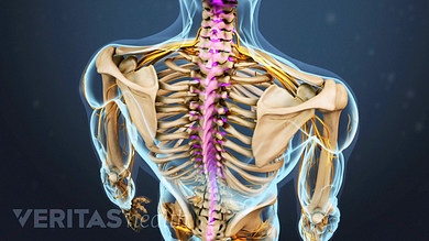
Your lower back (lumbar spine) is the anatomic region between your lowest rib and the upper part of the buttock. The rib cage also anchors the bones of the head, neck, shoulders, and arms to the trunk of the body. The cervical spine protects the nerves connecting to. Both the deltoid and the trapezius are firmly attached to the spine of the scapula. The trapezius and latissimus dorsi muscles connect the upper limb to the vertebral column.
Diagram Of Muscles In Upper Back Muscle Transparent Png 600x400 Free Download On Nicepng from www.nicepng.com The cause may be poor posture (such as forward head posture) or any type of irritation of the large back and shoulder muscles, including muscle strain or spasms. They originate from the vertebrae and insert into the scapulae. Learn to draw the upper back muscles by understanding the anatomical details and forms. The main superficial muscles of the back are the following: Anatomy of the upper back. The seventh cervical vertebra, referred to as c7, meets the first of 12 thoracic vertebrae t1 at the base of the neck, a. There are several different layers of muscles in your back. The bones of the chest and upper back combine to form the strong, protective rib cage around the vital thoracic organs such as the heart and lungs.
Balance the weight of your head on top of your spine.
In the upper back region, the trapezius, rhomboid major, and levator scapulae muscles anchor the scapula and clavicle to the spines of several vertebrae and the occipital bone of the skull. Balance the weight of your head on top of your spine. The cervical spine supports the weight and movement of your head and protects the nerves exiting your brain. Human musculature bodybuilding infographic muscular system vector human anatomy back muscle anatomy bicep male muscular anatomy human body anatomy female female anatomy muscle hamstrings muscle. Human anatomy · july 23, 2016. Related posts of upper back muscle diagram muscle anatomy basics. The traps) the latissimus dorsi (a.k.a. Anatomy of the upper back muscles. These muscles give height and breadth to back development. Powerful muscles that move the head and arms attach to these bones as well. This muscle is located on the upper portion of the back anatomy, underneath the trapezius. Musculoskeletal, shoulder & back back muscles, shoulder muscles. The iliocostalis muscles are furthest from the spine.
The trapezius and latissimus dorsi muscles connect the upper limb to the vertebral column. The superficial back muscles are situated underneath the skin and superficial fascia. It consists of seven vertebrae. The trapezius and latissimus dorsi muscles connect the upper limb to the vertebral column. It is like that for several reasons, all of which you can understand by looking at the anatomy of the thoracic spine.
How Upper Back And Shoulder Posture Influence Each Other And Cause Pain Oregon Exercise Therapy from www.oregonexercisetherapy.com Anatomy of the back organs. The back is the body region between the neck and the gluteal regions. Before giving our recommendations for upper back exercises, it's important to first go over the anatomy of the back musculature. The basic anatomy of your upper back by lindsey mcfadden as you're doing your regular upper back stretching exercises , you're probably wondering about the components of your upper back and why it happens to be the most stable part of your spine. Human anatomy · july 23, 2016. Your back consists of a complex array of bones, discs, nerves, joints, and muscles. The cervical section (the neck), the thoracic section (the upper back), the lumbar section (the lower back), the sacrum (part of the. The muscles of your back support your spine, attach your pelvis and shoulders to your trunk, and provide mobility and stability to your trunk and spine.
The rib cage also anchors the bones of the head, neck, shoulders, and arms to the trunk of the body.
The back is the body region between the neck and the gluteal regions. Learn to draw the upper back muscles by understanding the anatomical details and forms. Each block is separated by a disc that sits in between and each vertebra has a facet joint on either side. The iliocostalis muscles are furthest from the spine. The bones of the chest and upper back combine to form the strong, protective rib cage around the vital thoracic organs such as the heart and lungs. The nervous system of the thorax is a vital part of the nervous system as a whole, as it includes the spinal cord, peripheral nerves, and autonomic ganglia that communicate with and control many vital organs. One 2015 mayo clinic review of studies suggests that about a third of people get lower back or neck pain (a little higher for lower back, a little lower for neck), compared to less than one. There are several different layers of muscles in your back. This curve, called lordosis, helps to: It consists of seven vertebrae. The trapezius and latissimus dorsi muscles connect the upper limb to the vertebral column. The cause may be poor posture (such as forward head posture) or any type of irritation of the large back and shoulder muscles, including muscle strain or spasms. Back muscles anatomy here include the trapezius, latissimus dorsi, rhomboid and levator scapulae.
The back functions are many, such as to house and protect the spinal cord, hold the body and head upright, and adjust the movements of the upper and lower limbs. Muscles of the posterior neck and the back. This is my video about the muscles of the back. If you want to understand your upper back pain, start with an anatomy lesson. Musculoskeletal, shoulder & back back muscles, shoulder muscles.
Spinal Anatomy And Back Pain from embed.widencdn.net This muscle is located on the upper portion of the back anatomy, underneath the trapezius. Each block is separated by a disc that sits in between and each vertebra has a facet joint on either side. See back muscle anatomy stock video clips. It is very stiff, and the thoracic spine has a limited range of motion. They originate from the vertebrae and insert into the scapulae. Anatomy of the back organs. Both the deltoid and the trapezius are firmly attached to the spine of the scapula. Related posts of upper back muscle diagram muscle anatomy basics.
The trapezius and latissimus dorsi muscles connect the upper limb to the vertebral column.
Anatomy of the back organs. Both the deltoid and the trapezius are firmly attached to the spine of the scapula. Upper back pain is most commonly caused by muscle irritation or tension, also called myofascial pain. The cervical spine protects the nerves connecting to. These muscles give height and breadth to back development. This muscle is located on the upper portion of the back anatomy, underneath the trapezius. Learn to draw the upper back muscles by understanding the anatomical details and forms. The main superficial muscles of the back are the following: Human musculature bodybuilding infographic muscular system vector human anatomy back muscle anatomy bicep male muscular anatomy human body anatomy female female anatomy muscle hamstrings muscle. All about the back muscles function of the back muscles. This curve, called lordosis, helps to: Balance the weight of your head on top of your spine. There is a set of muscles in the upper back (called the thoracic area) called the spinalis thoracis.